3D printed microscope project progress update
Abstract
Many tasks in biology require tiny, accurate motion – achieved with expensive hardware. We have used inexpensive, 3D printed parts to make high performance mechanisms such as a compact mechanical stage, enabling an inexpensive microscope. This project aimed to improve the microscope (higher resolution imaging, fluorescence, phase contrast) and also to demonstrate other devices including a micro manipulator, suitable for use in electrophysiology for patch clamping or microinjection.
Goals
The project had the following goals for outcomes:
i) Improving microscope’s biological imaging capabilities (adding fluorescence and phase contrast)

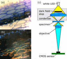
The basic configuration of our microscope uses transmission illumination: this gives bright-field images, suitable for observing many transparent samples. Adding a condenser lens in a printed holder allowed dark-field and basic phase contrast imaging, allowing a greater range of samples to be observed (See Figure 1).
The inverted design of the microscope means that samples are generally imaged through slides or coverslips. Inverted microscopes work well with a wide range of samples, including cell cultures and microtomed specimens.
Adding fluoresence imaging is still a work in progress
ii) Use in an incubator at the Light Microscopy Facility in the Cancer Research Institute.
iii) Plastic micromanipulators for microinjection or electrophysiology
iv) Use of ABS plastic as a potential replacement for PLA, as it has the potential to further improve the performance of printed mechanisms.
v) Science outreach kit
Progress and Outcomes
1. Open-source documentation
There are now full, illustrated assembly instructions available at docubricks as well as all source files available from GitHub. The GitHub repository contains the latest releases, in DocuBricks format, as well as the cutting-edge development options which include designs for use with RMS standard microscope objectives, and fluorescence filter mounts.
An extensive mechanical characterisation was performed by James Sharkey during his Part III project, which formed the basis of a publication describing the microscope’s mechanical design. This has been made open-access thanks to the SynBioFund grant.
2. Improved Imaging
The basic microscope design employs a webcam lens, taken from v1 of the Raspberry Pi camera module. v2 of the camera module already represents significantly improved resolution (by about 50%) due to its superior optics. However, for many scientific and medical applications it’s necessary to go beyond what is possible with a webcam lens. There is now a larger version of the microscope, capable of accepting an RMS standard objective (4x-100x have been tested, and work). This is used together with a 40mm planoconvex lens to correct for the shorter tube length and small sensor, to make the Raspberry Pi sensor cover the same field of view as a camera sensor 14.4x10.8mm in size (somewhere between a 1” and 4/3” video sensor) connected to a conventional lab microscope.
Fluorescence was demonstrated during Darryl Foo’s vacation project on the microscope, and has been re-integrated into the high-resolution objective-based design in a branch of the GitHub repository. This design will be used by the OpenPlant project “Establishing 3D Printed Microfluidics for Molecular Biology Workflows”.
Dark field and basic phase contrast imaging have been demonstrated in prototype designs, however we have not yet standardised and documented the build process for these. Both of these enhancements should be able to be made much more reproducible with our printed, adjustable condenser module which is present in the development branches on GitHub although not yet part of the documented releases.
3. Micromanipulation
A number of variations of the OpenFlexure microscope have been produced to give full 3D motion of an object. Initial designs proved insufficiently stiff, and suffered greatly from coupling between translational and rotational motion. One of these, a delta robot design, was improved and used by the 2015 Sensors CDT cohort as part of their group project, creating an Optical Projection Tomography system (http://cdt.sensors.cam.ac.uk/news/team-project-optical-projection-tomogr...).
More recently, a new variation on the design incorporating two-stage mechanical reduction has allowed 100nm-scale motion with very low wobble and good orthogonality between axes. It is currently designed in the form factor of a fibre-coupling stage, such as the flexure stages found in many optics laboratories that inspired the original flexure-based microscope. Initial testing suggests it is a very promising design, with sufficient accuracy and stability to be used in aligning single mode optical fibres. This design is on GitHub in development form, though it is yet to be packaged and documented for release.
Outreach
The primary impact of this project so far is the creation of WaterScope, a not-for-profit star-up aiming to use microscopy to bring about better diagnostics for public health and sanitation in the developing world. We are working together with makers in Tanzania (STICLab) and others affiliated with the Tech for Trade network, such as AB3D in Nairobi, to investigate the financial and technical feasibility of producing microscopes locally, freeing them from reliance on imports and the often unreliable supply chain that entails.
We have also worked with Public Lab, a US based organisation aiming to use the microscope as part of an air quality monitoring project. They have reproduced our high-resolution microscope, and are investigating creating a ruggedised case.
Finally, we have taken the microscope to various workshops and school visits, and have worked with a number of collaborators in Cambridge and beyond to test it out widely. We are currently refining the design and working to collate teaching materials around the microscope, which we aim to make available soon.
Expenditure
The initial £4000 budget was divided into:
£1400: open access publication fee for Review of Scientific Instruments paper
£1600: purchase of Ultimaker 2 3D printer (this enabled faster prototyping and had better axis orthogonality than the RepRap machine. It also made some successful ABS prints, and we intended to compare these to PLA ones particularly as regards stability at 37C. Unfortunately the ABS microscopes did not print as well so their performance couldn’t be meaningfully compared.)
£400: purchase of various optical components for testing purposes to achieve higher resolution imaging and fluorescence. Support in kind from WaterScope enabled sourcing of inexpensive objectives from Chinese manufacturers.
£600: consumables for constructing microscopes and testing them, including Raspberry Pis, Camera modules, plastic filament and nuts and bolts.
Plans for follow up
We intend to use the follow-on funding of £1000 to purchase the electronics and hardware to create a set of demo microscopes that might be lent to schools or other organisations in support of outreach or collaboration activities. This will fund the purchase of enough hardware to produce 5-10 microscopes, complete with Raspberry Pis and screens as necessary, which will make it much easier to take demonstrations into schools, etc.

