The aim of this project is to investigate if piezoelectric materials can be used to interface directly with biological systems by changing aspects of their mechanical environment. Piezoelectric materials can both detect and apply forces, therefore could be used to create some form of 'touch screen' for cells. Various aspects towards this aim have been investigated, including creating and characterising nanowires of a piezoelectric polymer: poly-L-lactic acid. Aerosol Jet Printing has been investigated as a possible fabrication route for the device, and a matrix addressing scheme has been developed for device control. A working prototype is currently being constructed. Results from the investigation into poly-L-lactic acid nanowires were presented at the Nanogenerators and Piezotronics conference (Rome, June 2016) and are currently being prepared for publication. The follow-on funding available for this project will allow for this manuscript to be published in an open access format.
Outcomes, Outputs and Progress
Biological systems are responsive not only to chemical changes, but also changes in their mechanical environment. This 'mechanobiology' is believed to regulate some important cell functions, such as how stem cells differentiate and the direction in which brain cells can grow. Understanding exactly how these mechanical cues can influence behaviour could lead to developments in regenerative medicine, for example.
The forces that occur between cells do so over very short length scales and are exceptionally small. In order to mechanically interface with these cellular systems, it is necessary to create a sensing element that is equally small and sensitive.
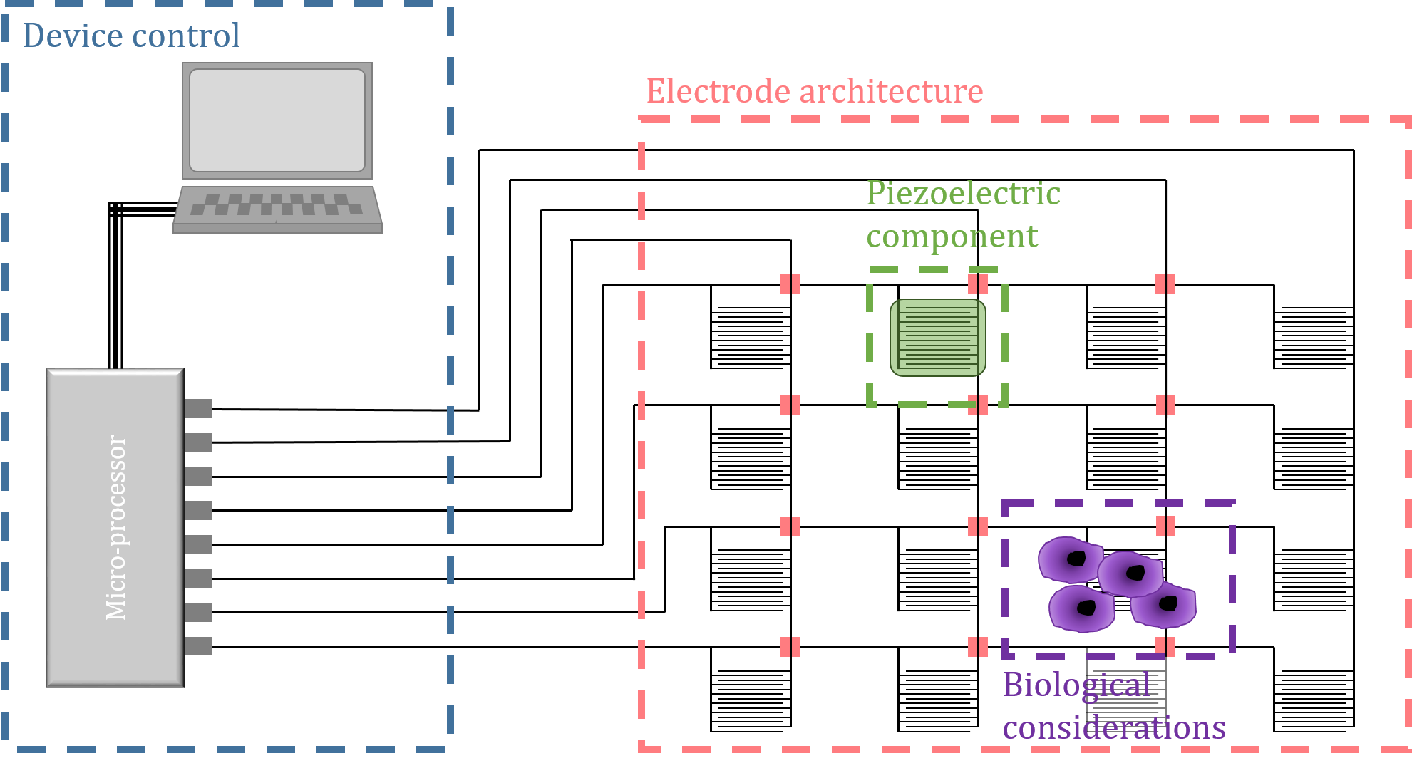
Nanowires of piezoelectric polymers are possible candidates for this. Piezoelectric materials can be used both to detect and apply forces, and by using a polymeric material and making the structures at the nanoscale, the sensitivity of the structures can be improved. The ultimate goal is to incorporate these nanowires into a grid of electrodes, allowing some spatial resolution to the measurements - therefore creating a touch screen for cells - as outlined in Figure 1. This was deemed a more appropriate name, rather than 'piezoelectric bioplatform' as was initially proposed.
The majority of the time on this project has been spent developing the active piezoelectric component of the proposed device, with the remainder of the time used to develop the interface electronics as well as possible manufacturing techniques. These elements are being brought together to create a working prototype.
Template grown poly-L-lactic acid nanowires for piezoelectric sensing and actuation
Poly-L-lactic acid (PLLA) is a piezoelectric polymer that is biologically derived, and as such inherently compatible with biological systems. PLLA was investigated for this project because of its biological nature, but also because the nature of its piezoelectricity is somewhat novel and is interesting to study in its own right.
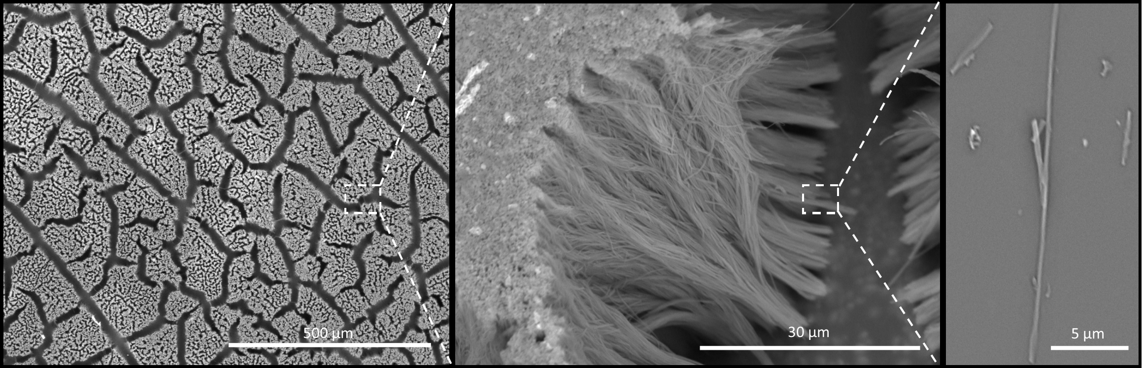
Nanostructures have previously been made from PLLA using a technique known as electrospinning. The equipment required for this technique is complex, expensive and was not available for this project. Instead, a more straightforward ‘template wetting’ method was developed. This involves infiltrating a nanoporous membrane (or ‘template’) with polymer solution. The pores in these membranes, which are used commercially as particulate filters, are around 200 nm in diameter and the membranes themselves are nominally 60 μm thick. Once the solution has crystallised, the template material can be etched away to reveal polymer nanowires with the same dimensions as the pores. This method has been used extensively for creating nanowires of other piezoelectric polymers, but not before been used successfully with PLLA. Scanning Electron Microscopy (SEM) images of the nanowires at various points in the growth process are shown in Figure 2.
It was found that changing some parameters of the growth process can influence aspects of the nanowire crystal structure. In order to optimise the sensing performance of the nanowires, it seemed reasonable to tune these growth conditions to target the crystal structure with strongest piezoelectric output. However, whilst PLLA is recognised as a piezoelectric polymer, the crystallographic origins of piezoelectricity in this material are not that well understood. The crystal structure of PLLA has been extensively studied, but never in the context of its piezoelectric properties. Therefore it was not possible to explicitly state which crystal phase, and thus which growth conditions, should be used.
In order to inform the choice of growth conditions, the piezoelectric properties of the nanowires had to be investigated and correlated with characterisation of the material’s structural. The intention was to answer the following questions:
-
Of the 4 crystal phases of PLLA, which (if any) is responsible for its piezoelectric properties
-
To what extent is alignment of crystallites and/or non-crystalline polymer chains important for revealing piezoelectricity?
-
Does the template wetting method favour the formation of a particular crystal phase?
-
Does the template wetting method lead to any preferential alignment of the polymer, either as crystallites or polymer chains?
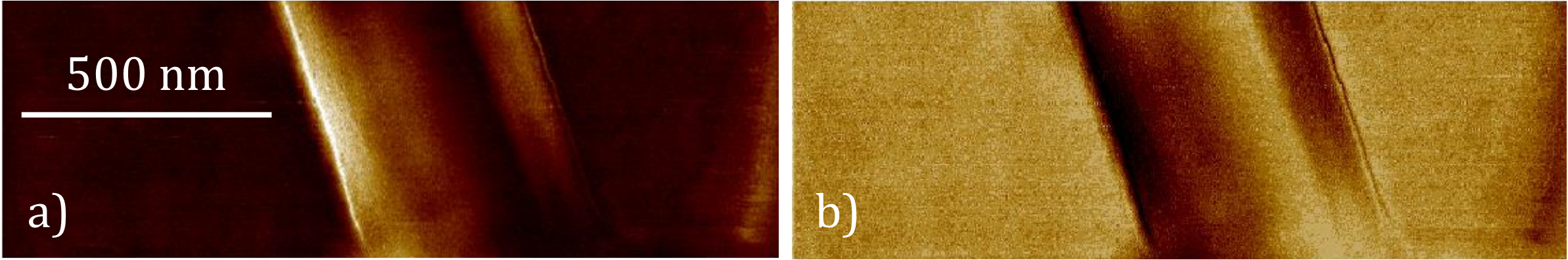
Given the nature of piezoelectricity in PLLA, Piezo-Force Microscopy (PFM) was used to probe the piezoelectric response of individual nanowires, as shown in Figure 3. Achieving quantitative results from PFM images of nanowires is challenging, especially in the case of PLLA given its specific piezoelectric properties. Characterising the piezoelectric properties of PLLA nanowires using this technique has taken longer and proved more challenging than expected, which has delayed the progress of the project somewhat. However, the characterisation of PLLA nanowires in this manner has not before been achieved and has proved to be exceptionally informative.
X-ray diffraction (XRD) and Differential Scanning Calorimetry (DSC) have been used to characterise the structure of the nanowires. The results from the structural and piezoelectric characterisation can then be compared to reveal if any correlation exists. The results of this investigation were presented at a recent conference (NGPT - Rome, June 2016) and the slides from this investigation are freely available, attached to this report. A manuscript detailing the outcomes of this investigation is also under preparation. It is hoped that using the follow on funding available from the SynBio fund that the results can be published in an open-access journal.
Biological aspects
The motivation of this project is inherently biological, therefore some consideration towards this must be made. Initially, there were ideas to use E. coli bacteria as test cells for the prototype. However, as the length scales that might be easily achievable with respect to device fabrication became apparent, it seemed reasonable to choose a larger cell line.
We approached the Cambridge Centre for Medical Materials here in the Materials Science department and discussed the possibility of using some mammalian cells. Fibrosarcoma cells were suggested, which are cells taken from a fibrous tumour and are known to exert a reasonable degree of traction force whilst being relatively straightforward to culture \cite{Rasheed1974}.
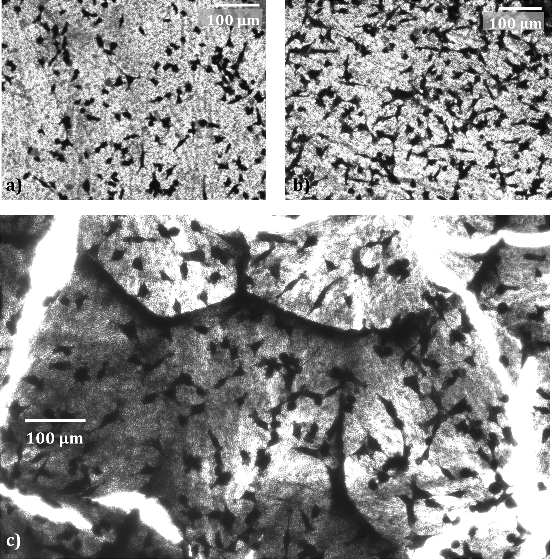
Poly-L-lactic acid is widely used in biomedical applications and its biocompatibility is broadly accepted. However, the term ‘biocompatible’ is rather broad and extends beyond intrinsic material properties; the morphology of a material is also very important. Therefore, in order to test that the PLLA nanowires created here are indeed suitable, fibrosarcoma cells were grown on the samples produced. This was only a simple test, but initial results seemed promising with the cells able to survive and adhere to the nanowire structure.
The cells were imaged optically, as shown in Figure 4, however the topography of the nanowire samples meant that the images obtained from this investigation technique were of reasonably low quality. An alternative imaging method must therefore be used. The possibility of using fluorescence microscopy is currently being investigated. Another alternative is to dehydrate the cells and observe the samples using Scanning Electron Microscopy (SEM).
Interface electronics
In order to simplify the project, it was decided to initially focus only on applying signals to the piezoelectric material. In order to achieve some spatial resolution to the applied signals, an array of ‘pixels’ is required. Each pixel should be individually addressable so that the physical stimulation can be targeted and varied across the device.
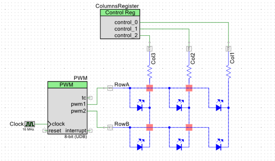
A matrix addressing scheme was developed to achieve this. This method allows for an array of m x n elements to be controlled using m + n electrodes and is very similar to the scheme used in smartphone touchscreens and LCD displays. A test circuit with LEDs at each pixel was constructed around a micro-processor development board (Sparkfun FreeSOC2 Development Board – Cypress PSoC5LP chip). A schematic of the circuit is shown in Figure 5. This allowed for the brightness and breathing rate of 6 LEDs to be controlled independently using just 5 electrodes. The source code used for this is available in the attached main.c file. The aim is to transfer this to a working prototype of the device, with piezoelectric material replacing the LED at each pixel.
Fabrication methods
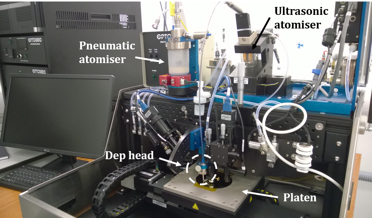
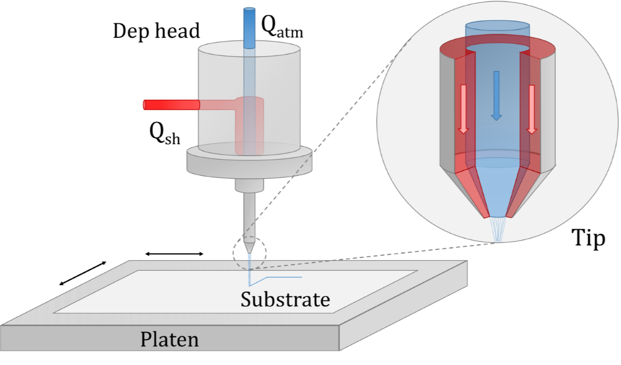
The uses of Aerosol Jet Printing (AJP) within this device have also been investigated. AJP is an additive manufacturing technique that can be used for wide-area deposition of fine-scaled features. It is typically used to pattern circuits in consumer electronics, and therefore could be used to print the electrodes required for this device. Since the system installed in the department – see Figure 6 - is relatively new, much work has had to be carried out to optimise and understand the deposition characteristics.
Investigation has also been carried out into producing polymer inks that could form a 'functional' piezoelectric ink. This could replace or perhaps augment the nanowires outlined above as the active piezoelectric component. Achieving a stable ink with appropriate viscosity is a significant challenge here, but work is progressing steadily.
Expenditure
A brief overview of how the money was spent is given in Table 1, although a more detailed breakdown of each item is also available.
| Expenditure | Amount (£) |
| Lab consumables | 96.07 |
| Chemicals and materials | 1853.99 |
| Research equipment time | 125.63 |
| PFM tips | 1508.00 |
| Travel | 405.26 |
| Total | 3988.95 |
One of the most significant costs in this project was the PFM tips. These are similar to standard AFM tips, but must be doped to make them electrically conducting. Furthermore, the stiffness of these cantilevers must be optimised to probe the properties of polymers, which are generally reasonably compliant.
The travel funding allowed attendence to an Aerosol Jet Printing user group meeting hosted by Optomec, the manufacturers of the system in the Materials Science department. Given our relative inexperience with this technology, the meeting proved invaluable for learning new techniques and information regarding ink formulation.
Request and justification for follow on funding
We would like to request the additional funding available for this project. A manuscript detailing the findings of the poly-l-lactic acid nanowire investigation is currently being prepared and the follow on funding will allow for this to be published in an open access journal. It is hoped that these results will help to clarify the origins of the piezoelectric effect in this material. We do acknowledge that the aims of this investigation are mainly concerned with fundamental material properties, rather than synthetic biology, however the relevance of poly-l-lactic acid within biology – both in the context of this project but also more generally for biomedical implants – means that PLLA and its properties are worth investigating.
Conclusion
While a working protoype has yet to be achieved, key milestones towards this goal have been reached. This includes material fabrication and characterisation, biological and electronic interfacing, and optimising the device design. The outcomes of the material investigation have been presented at a recent conference (Nanogenerators and Piezotronics - NGPT - Rome, June 2016) and a manuscript detailing the results is currently being prepared. The follow-on funding available for this project will allow for these results to be presented in an open access journal.
The various aspects of the project are now at a stage where they can begin to be brought together. Whilst there will inevitably be future challenges to overcome, we are confident that a working device can be produced soon.